One flu east, one flu westThe
outbreak of a new flu strain—a nasty mash-up of swine, avian, and
human viruses—has infected 1,000 people in Mexico and the U.S.,
killing 68. The World Health Organization warned Saturday that the outbreak could reach global pandemic levels.
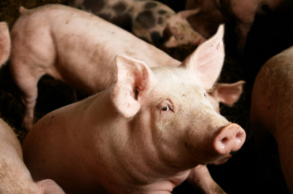
Is Smithfield Foods, the world’s largest pork packer and hog producer, linked to the outbreak? Smithfield operates massive hog-raising operations Perote, Mexico, in the state of Vera Cruz, where the outbreak originated. The operations, grouped under a Smithfield subsidiary called Granjas Carroll, raise 950,000 hogs per year, according to the company Web site.
On Friday, the U.S. disease-tracking blog Biosurveillance published a timeline of the outbreak containing this nugget, dated April 6 (major tip of the hat to Paula Hay, who alerted me to the Smithfield link on the Comfood listserv and has written about it on her blog, Peak Oil Entrepreneur):
Residents [of Perote] believed the outbreak had been caused by contamination from pig breeding farms located in the area. They believed that the farms, operated by Granjas Carroll, polluted the atmosphere and local water bodies, which in turn led to the disease outbreak. According to residents, the company denied responsibility for the outbreak and attributed the cases to “flu.” However, a municipal health official stated that preliminary investigations indicated that the disease vector was a type of fly that reproduces in pig waste and that the outbreak was linked to the pig farms. It was unclear whether health officials had identified a suspected pathogen responsible for this outbreak.
From what I can tell, the possible link to Smithfield has not been reported in the U.S. press. Searches of Google News and the websites of the New York Times, Washington Post, and Wall Street Journal all came up empty. The link is being made in the Mexican media, however. “Granjas Carroll, causa de epidemia en La Gloria,” declared a headline in the Vera Cruz-based paper La Marcha. No need to translate that, except to point out that La Gloria is the village where the outbreak seems to have started. Judging from the article, Mexican authorities treat hog CAFOs with just as much if not more indulgence than their peers north of the border, to the detriment of surrounding communities and the general public health. Get this:
De acuerdo con uno de los habitantes de la comunidad, Eli Ferrer Cortés, los desechos fecales y orgánicos que produce Granjas Carroll no son tratados adecuadamente, lo que genera contaminación del agua y del viento en la region.
My rough translation: According to one community resident, the organic and fecal waste produced by Granjas Carrol isn’t adequately treated, creating water and air pollution in the region. I witnessed—and smelled—the same thing in Hardin County, Iowa, a couple of years ago, another area marked by intensive industrial hog production. The article goes on to say that area residents have long complained of “fetid odors” in the air and water, and swarms of flies hovering around waste lagoons. Like their counterparts who live in CAFO-heavy U.S. areas, they also complain of respiratory ailments. Now, with 30 percent of the area’s residents now infected with the virulent flu bug, people are demanding that state and federal authorities inspect hog operations there. So far, reports La Marcha, the response has been: nada.
The Mexico City daily La Jornada has also made the link. According to the newspaper, the Mexican health agency IMSS has acknowledged that the orginal carrier for the flu could be the “clouds of flies” that multiply in the Smithfield subsidiary’s manure lagoons.
I’ll be in touch with contacts in Mexico as this story develops —and I’ll be curious to see whether the U.S. media explores the link with Smithfield’s Mexico operation.
Note: In the original version of this post, I had called production at Granjas Carroll “nearly equal to Smithfield’s total U.S. production.” I had been confusing total production at Granjas Carroll—950,000 hogs produced in fiscal 2008—with the number of sows, or breeding pigs, kept by Smithfield in the United States. According to my source—“Concentration of Ag Markets, 2007” (PDF) by Hendrickson and Heffernan—Smithfield keeps 1.2 million sows. Actual hog production is much larger—thus Smithfield’s total U.S. hog production is much larger than Granjas Carroll’s. I regret the error.
Comments
View as Flat
Zana Posted 8:34 pm
25 Apr 2009
Permalink
pioneer989 Posted 8:55 pm
25 Apr 2009
Permalink
AngieWalsch Posted 12:21 am
26 Apr 2009
Permalink
Roysc Posted 5:19 am
26 Apr 2009
Permalink
Bud Dingler Posted 9:50 am
26 Apr 2009
You know darned well your claims are not substantiated by any facts. This kind of hype puts a bad name to Grist.This site says it well
http://www.fairfoodfight.com/blog/el-dragón/grist-cafos-blame-h1n1-swine-flu-reallyFlu pandemics existed long before feedlots became the norm.
Permalink
Tom Philpott Posted 10:38 am
26 Apr 2009
del Seguro Social (IMSS), el vector epidémico serían las nubes de
moscas que despiden las granjas porcícolas y las lagunas de oxidación
donde la empresa mexicana-estadunidense arroja toneladas de estiércol.Rough translation: "According to physicians from the state of Vera Cruz and the Mexican health agency IMSS, the epidemic's vector could be the clouds of flies created by hog CAFOs and the manure lagoons where the U.S.-Mexican company (Smithfield's Granjas Carroll subsidiary) through tons of manure."Don't you think it's news that the Mexican healthcare ministry is looking at U.S.-owned CAFOs as the source of a global flu pandemic? Yes, there were flu pandemics before CAFOs, just as there were wars before the advent of fighter planes and bombs. Things have changed a bit since, haven't they?
Permalink
enviroperk Posted 8:36 am
27 Apr 2009
Permalink
guanoguy Posted 1:13 pm
26 Apr 2009
http://www.newscientist.com/article/dn17026-swine-flu-what-you-need-to-know.html
"Does this virus mean I shouldn't eat pork?
No. This virus is named swine flu because one of its surface proteins is most similar to viruses that usually infect pigs. But we've never seen this particular virus in pigs before. It is spreading in people; that's the problem."So I gather that there really is no connection to the Swine flu outbreak and any farms with pigs on them.
Permalink
PurpleOzone Posted 3:16 pm
26 Apr 2009
One woman masquerading as a reporter asked in Obama "has been vaccinated with Tamiflu & Renazen" (I forget the exact name of the other antiviral drug useful with this flu.) The CDC guy explained her 'misconception' and answered 'we don't have vaccines against a new flu'. A pretty young woman asked repeatedly if the flu is 'bioterrorism'. Darn it, the doctor couldn't agree, so there goes her chance for prime time.
Memo to TV news shows, newspapers, magazines, etc: Could you please verify that a 'reporter' has an IQ above 85 before you issue a press card? These cretins soak up air time which could be used for info the public needs.
Permalink
Erik Hoffner Posted 3:24 pm
26 Apr 2009
Erik, Orion Grassroots Network
Permalink
guanoguy Posted 6:28 pm
26 Apr 2009
Permalink
PHEP Professional Posted 7:04 pm
26 Apr 2009
Permalink
Erik Hoffner Posted 7:13 am
27 Apr 2009
when a young woman tested positive for a new strain of MRSA, called
ST398. The family lived on a farm, so public health authorities swept
in — and found that three family members, three co-workers and 8 of 10
pigs tested all carried MRSA.""Now this same strain of MRSA has also been found in the United States. A new study by Tara Smith, a University of Iowa epidemiologist, found that 45
percent of pig farmers she sampled carried MRSA, as did 49 percent of
the hogs tested."These quotes are from a piece in a recent edition of the New York Times. No doubt you have a 'professional' take on this.Erik, Orion Grassroots Network
Permalink
Bud Dingler Posted 4:48 pm
26 Apr 2009
Permalink
PHEP Professional Posted 7:03 pm
26 Apr 2009
Permalink
amazingdrx Posted 7:18 pm
26 Apr 2009
Permalink
MichiganGrassFarmer Posted 6:16 am
27 Apr 2009
For those following the swine flu story concerned about a possible pandemic, you should be concerned about a program being fought by many small farmers in the US right now that can have a direct affect on your family's health. The program, National Animal Identification System (NAIS), falsely labeled as a disease control program, is designed to help big agribusiness at the expense of small farmers and every person who owns even one animal.
It is hard to believe that most people still don't know that factory farms concentrating animals in confinement spread diseases while small family farms with animals on grass in the sunshine minimize diseases by allowing manure to be spread out in small amounts quickly utilized by the soil. What kind of farm would you rather live next to? NAIS will encourage the wrong kind of farms while shutting down the right kind.
NAIS takes agriculture in the wrong direction, against nature. But remember, nature always bats last. Allowing NAIS to go through will enable the factory farms with manure lagoons breeding the next pandemic, exposing millions to increased risk of death from disease. Contact your representatives and tell them to stop NAIS. (now watch the apologists for factory farms and proponents of giving up your rights come out of the woodwork) Remember, an eartag never stopped a disease.
Mike Murphy
Michigan
Permalink
PatCullen Posted 7:47 am
27 Apr 2009
Permalink
kristennicole Posted 9:33 am
27 Apr 2009
Permalink
hutena Posted 2:11 pm
27 Apr 2009
British scientist, Terry Mabbett, reporting in a recent issue of Poultry International, says the new research findings must come as a big wake up call for the world's poultry industry.
"That the avian influenza (AI) virus can be spread by winged insects as well as wild birds underlines the need for efficient fly control on poultry farms along with other strict biosecurity measures," he says.
According to Mabbett, studies recently carried out at the North Carolina State University, adult houseflies were seen to carry infectious doses of the Newcaslte Disease Virus in their guts for up to three hours after feeding. This he says, "might be important for the spread of the virus when fly populations are high and in contact with highly virulent NDV strains." The scientist has also cited earlier reported instances of houseflies carrying avian influenza virus. A 1985 study based on a serious 1983/84 outbreak of H5N2 in Lancaster County Pennsylvania (USA) where nearly 90 per cent of the affected poultry stocks died.
More than a third of the housefly samples collected from the vicinity of the outbreak contained bird flu virus particles.
Similarly, blow flies caught near a Kyoto poultry farm in Western Japan following an H5N1 outbreak in 2004 also carried doses of the virus. Dr Mabbett says the presence of avian influenza virus in Musca domestica or other flies has opened up a whole new dimension on this virus disease.
Indian poultry experts told Asian Age that "at the very minimum, poultry farm owners need to put their house in order. Our poultry farms could be particularly susceptible to insect-borne transmissions of the virus because of the abysmal sanitation maintained."
Permalink
enviroperk Posted 2:42 pm
27 Apr 2009
Permalink
sindark Posted 2:59 pm
27 Apr 2009
I am not saying this swine flu is definitely the product of factory farming: just that it is plausible and worth investigating.
Permalink
Green Granny Posted 6:06 pm
27 Apr 2009
I sincerely hope this "outbreak" is contained. Every time I turn on the radio, I'm told it has spread to yet another country/city.
Permalink
mexture Posted 6:45 pm
27 Apr 2009
Permalink
enviroperk Posted 7:38 pm
27 Apr 2009
Permalink
RandyT Posted 6:54 am
29 Apr 2009
Permalink
mexture Posted 7:47 pm
27 Apr 2009
Permalink
ferrand Posted 7:12 am
28 Apr 2009
www.hm-treasury.gov.uk/d/climatechange_grunhausproject.pdf
Permalink
rmk-nj Posted 8:12 am
28 Apr 2009
"In Mexico, state health authorities looking for the initial source of the outbreak toured a million-pig hog farm in Perote, in Veracruz State. The plant is half-owned by Smithfield Foods, an American company and the world’s largest pork producer. Mexico’s first known swine flu case, which was later confirmed, was from Perote, according to Health Minister José Ángel Córdova. The case involved a 5-year-old boy who recovered.
But a spokesman for the plant said the boy was not related to a plant worker, that none of its workers were sick and that its hogs were vaccinated against flu."
Permalink
amazingdrx Posted 8:46 am
28 Apr 2009
Permalink
amazingdrx Posted 9:18 am
28 Apr 2009
Permalink
amazingdrx Posted 9:22 am
28 Apr 2009
Permalink
Erik Hoffner Posted 9:27 am
28 Apr 2009
But Jose Luis Martinez, a 34-year-old resident of La Gloria, said he knew the minute he learned about the outbreak on the news and heard a description of the symptoms: fever, coughing, joint aches, severe headache and, in some cases, vomiting and diarrhea.
"When we saw it on the television, we said to ourselves, 'This is what we had,'" he said Monday. "It all came from here. ... The symptoms they are suffering are the same that we had here." "http://news.yahoo.com/s/ap/20090428/ap_on_re_la_am_ca/lt_swine_flu_mexico_ground_zero
Permalink
eeaaddgg Posted 11:08 am
28 Apr 2009
Permalink
enviroperk Posted 3:01 pm
28 Apr 2009
Permalink
kenspear Posted 3:08 am
29 Apr 2009
Permalink
Missouri Mama Posted 6:36 am
29 Apr 2009
Permalink
Former Ag Teacher Posted 7:39 pm
29 Apr 2009
Permalink
Bud Dingler Posted 10:22 am
29 Apr 2009
Who knew that the COP right wing wackos had any friends on the other end of the political spectrum?
Permalink
atreyger Posted 10:41 am
29 Apr 2009
Permalink
enviroperk Posted 11:11 am
29 Apr 2009
Permalink
Tom Philpott Posted 11:59 am
29 Apr 2009
international team of scientists—including Jay Graham and Ellen Silbergeld of Johns
Hopkins—published in the May-June 2008 Public Health Reports, entitled “The
Animal-Human Interface and Infectious Disease in Industrial Food Animal
Production: Rethinking Biosecurity and Biocontainment” (PDF)An analysis of data from the Thai government investigation in 2004 indicates that the odds of H5N1 outbreaks and
infections were significantly higher in large-scale commercial poultry operations as compared with backyard flocks. These data suggest that successful strategies to prevent or mitigate the emergence of pandemic avian influenza must consider risk factors specific to modern industrialized food animal production.
Permalink
RandyT Posted 10:55 am
29 Apr 2009
Permalink
enviroperk Posted 12:31 pm
29 Apr 2009
Permalink
ejustice Posted 12:49 pm
29 Apr 2009
Permalink
RandyT Posted 1:01 pm
29 Apr 2009
Permalink
gaspard.leon Posted 6:27 pm
29 Apr 2009
What's most likely on the lack of evidence front is that the commercial farm opererators and owners don't have much incentive to do studies or let studies be done on their farms, so there is not much data around.
Interesting reading, a good summarization of the commercial farming problem:
http://grain.org/articles/?id=48
A quote from the story above: "It should be noted that a common ingredient in industrial animal feed is "poultry litter", which is a mixture of everything found on the floor of factory poultry farms: fecal matter, feathers, bedding, etc"
good way to mix pig and bird flu strains?
Permalink
Former Ag Teacher Posted 7:58 pm
29 Apr 2009
Permalink
gristle Posted 1:56 am
04 May 2009
Critics: US Doing Too Little to Prevent 'Mad Cow'
by Libby Quaid
http://www.commondreams.org/headlines05/0618-02.htmWASHINGTON -- American cattle are eating chicken litter, cattle blood and restaurant leftovers that could help transmit mad cow disease -- a gap in the U.S. defense that the Bush administration promised to close nearly 18 months ago."Once the cameras were turned off and the media coverage dissipated, then it's been business as usual, no real reform, just keep feeding slaughterhouse waste," said John Stauber, an activist and co-author of Mad Cow USA: Could the Nightmare Happen Here?He contended, "The entire U.S. policy is designed to protect the livestock industry's access to slaughterhouse waste as cheap feed."Loopholes in Ban on Cattle Remains in FeedThe Food and Drug Administration promised in January 2004 to close loopholes in a ban on putting cattle remains in cattle feed, but it has failed to act. The government calls the ban a "firewall" against the spread of mad cow disease. Eating the mad cow disease protein is the only way cows are known to get the disease.The Food and Drug Administration promised to tighten feed rules shortly after the first case of mad cow disease was confirmed in the U.S., in a Washington state cow in December 2003.
"Today we are bolstering our BSE firewalls to protect the public," Mark McClellan, then-FDA commissioner, said on Jan. 26, 2004. The FDA said it would ban blood, poultry litter and restaurant plate waste from cattle feed and require feed mills to use separate equipment to make cattle feed.However, last July, the FDA scrapped those restrictions. McClellan's replacement, Lester Crawford, said an international team of experts assembled by the Agriculture Department was calling for even stronger rules and that the FDA would produce new restrictions in line with those recommendations.Today, the FDA still has not done what it promised to do. The agency declined interviews, saying in a statement only that there is no timeline for new restrictions."It's just a lot of talk," said Rep. Rosa DeLauro, D-Conn., a senior House Democrat on food and farm issues. "It's a lot of talk, a lot of press releases, and no action." No reason to think that Mexico has done what the United States hasn't done either.
Permalink
RandyT Posted 6:19 am
30 Apr 2009
Permalink
Former Ag Teacher Posted 8:13 am
30 Apr 2009
Permalink
RandyT Posted 9:30 am
30 Apr 2009
(letter to Col. William F. Elkins)
Ref: The Lincoln Encyclopedia, Archer H. Shaw (Macmillan, 1950, NY) http://www.ratical.org/corporations/Lincoln.html As to how this flu can be passed on:"How does swine flu spread?
Influenza viruses can be directly transmitted from pigs to people and from people to pigs. Human infection with flu viruses from pigs are most likely to occur when people are in close proximity to infected pigs, such as in pig barns and livestock exhibits housing pigs at fairs. Human-to-human transmission of swine flu can also occur. This is thought to occur in the same way as seasonal flu occurs in people, which is mainly person-to-person transmission through coughing or sneezing of people infected with the influenza virus. People may become infected by touching something with flu viruses on it and then touching their mouth or nose." http://www.cdc.gov/swineflu/key_facts.htmSo back to the very strong possibility of these corporate hog farms being the source of this current outbreak, it is more than possible. For the interference of corporations into our lives for their profits I believe Lincoln was right on - and that is proven every day from these international factory farms to Wall Street "corporate" bankers and their money influence on our government and it's agencies.For some more detailed research into these corporate farms Pew Research conducted a very lengthy investigation into their effects, both in terms of health issues and actual economics. Final Report: Putting Meat on The Table: Industrial Farm Animal Production in Americahttp://ncifap.org/
Permalink
enviroperk Posted 9:53 am
30 Apr 2009
interview that pigs at the farm are from North America, while the
genetic material in the virus is from Europe and Asia."http://online.wsj.com/article/SB124105320874371313.htmlThe Lincoln quote is also hogwash (couldn't resist).http://www.snopes.com/quotes/lincoln.aspThough, I agree fully with the evils of most corporate livestock farming.
Permalink
gristle Posted 2:49 am
04 May 2009
http://www.usatoday.com/news/health/2009-05-03-canada-flu_N.htm Funny how that happens.
Permalink
Former Ag Teacher Posted 6:50 pm
30 Apr 2009
Permalink
enviroperk Posted 10:33 pm
30 Apr 2009
Permalink
RandyT Posted 9:14 am
05 May 2009
Permalink
moleculeman Posted 11:45 am
30 Apr 2009
Published online: 22 February 2009 | doi:10.1038/nsmb.1566Structural and functional bases for broad-spectrum neutralization of avian and human influenza A virusesJianhua Sui1,4, William C Hwang2,4, Sandra Perez3, Ge Wei2, Daniel Aird1, Li-mei Chen3, Eugenio Santelli2, Boguslaw Stec2, Greg Cadwell2, Maryam Ali1, Hongquan Wan3, Akikazu Murakami1, Anuradha Yammanuru1, Thomas Han1, Nancy J Cox3, Laurie A Bankston2, Ruben O Donis3, Robert C Liddington2 & Wayne A Marasco1AbstractInfluenza virus remains a serious health threat, owing to its ability to evade immune surveillance through rapid genetic drift and reassortment. Here we used a human non-immune antibody phage-display library and the H5 hemagglutinin ectodomain to select ten neutralizing antibodies (nAbs) that were effective against all group 1 influenza viruses tested, including H5N1 'bird flu' and the H1N1 'Spanish flu'. The crystal structure of one such nAb bound to H5 shows that it blocks infection by inserting its heavy chain into a conserved pocket in the stem region, thus preventing membrane fusion. Nine of the nAbs employ the germline gene VH1-69, and all seem to use the same neutralizing mechanism. Our data further suggest that this region is recalcitrant to neutralization escape and that nAb-based immunotherapy is a promising strategy for broad-spectrum protection against seasonal and pandemic influenza viruses.IntroductionSeasonal influenza A is a scourge of the young and old, killing more than 250,000 worldwide each year, while creating an economic burden for millions1. Pandemic influenza, which occurs when a new virus emerges and infects people globally that have little or no immunity, represents a grave threat to human health; for example, the 1918 Spanish Flu pandemic caused an estimated 50 million deaths2, 3. Vaccines have historically been the mainstay of infection control. However, owing to rapid antigenic drift, the vaccine antigen must be updated annually based on global influenza surveillance4, 5, and it is not always fully successful. In addition, some recent H5N1 vaccines have shown promising results6, 7, 8, 9, but none has been reported to elicit a broad neutralizing response in humans. Neuraminidase inhibitors, especially oseltamavir (Tamiflu), remain the primary treatment, but they have limited efficacy if administered late in infection, and widespread use is likely to result in the emergence of resistant viral strains10, 11.Influenza A is subclassified by its two major surface proteins: hemagglutinin, which mediates cell entry, first by recognizing host proteins bearing sialic acid on their surface, and second by triggering the fusion of viral and host membranes following endocytosis, allowing viral RNA to enter the cytoplasm; and neuraminidase, which cleaves sialic acid from host and viral proteins, facilitating cell exit12. There are 16 hemagglutinin subtypes (H1–16) and 9 neuraminidase subtypes (N1–9) that make up all known strains of influenza A viruses by various combinations of hemagglutinin and neuraminidase12 (Supplementary Fig. 1 online).The recent spread of highly pathogenic avian influenza (HPAI), caused by the H5N1 strain, across Asia, Europe and Africa raises the specter of a new pandemic, should the virus mutate to become readily transmissible from person to person. The evolution of H5N1 into a pandemic threat could occur through a single reassortment of its segmented genome or through the slower process of genetic drift12, 13. Nearly 400 human H5N1 infections have been reported since 1997 from 14 countries, with a case mortality rate in the immunocompetent population above 60%4.New therapeutic strategies that provide potent and broadly cross-protective host immunity are therefore a global public health priority. Human mAb-based 'passive' immunotherapy is now being used to treat numerous human diseases, including respiratory syncytial virus infection, and we have proposed how immunotherapy could be used strategically in a viral outbreak setting14.In the present study, we first used a phage-display antibody library and recombinant H5 trimeric ectodomain to isolate a group of high-affinity nAbs that were potent inhibitors of H5N1 viral infection in vitro and in vivo. On the basis of crystallographic and functional studies, we showed that the nAbs bind to a common epitope—a highly conserved pocket in the stem region of hemagglutinin containing the 'fusion peptide'—that rationalizes their ability to block membrane fusion rather than cell attachment. Sequence and structural analysis of all 16 hemagglutinin subtypes point to the existence of just two variants of this epitope, corresponding to the two classic phylogenetic groupings of hemagglutinin (groups 1 and 2). We therefore tested eight more group 1 hemagglutinin subtypes and demonstrated an unprecedented cross-subtype binding and/or neutralization spectrum. Because we had used a group 1 subtype (H5) for our panning, our nAbs, as expected, failed to neutralize group 2 subtypes H3 and H7. These results nevertheless raise the possibility that a cocktail comprising a small subset of nAbs raised against representatives of the two groups could provide broad protection against all seasonal and pandemic influenza A viruses.Top of pageResultsIdentification of nAbs against H5N1The current H5N1 epidemics involve viruses derived from a single lineage of H5 hemagglutinin. Within this lineage, four distinct clades have been identified as major threats to public health15, 16. We expressed recombinant trimeric ectodomain of H5 hemagglutinin from one of these viruses (strain A/Vietnam/1203/04 (H5N1), 'H5-VN04', clade 1) in insect cells17 (Supplementary Fig. 2 online), immobilized it on a plastic surface and selected antibodies from a 'non-immune' human antibody phage-display library (using single-chain VH-VL fragments ('scFv'))18. Two rounds of panning and the screening of 392 clones identified 10 unique antibodies formed by six distinct VH (variable region of heavy chain) fragments in combination with ten different VL (variable region of light chain) fragments (Supplementary Table 1 online).We found that all ten nAbs bound trimeric H5-VN04 with similar avidity, but did not bind monomeric HA1 (Fig. 1a). Presented as scFv-Fc constructs, they potently neutralized the clade 1 H5 pseudovirus, A/Thailand/2-SP-33/2004 (H5N1) ('H5-TH04') (Fig. 1b); and, in a stringent plaque-reduction assay, they all showed high levels of neutralization against H5-VN04, as well as the more divergent (clade 2.1) A/Indonesia/5/2005 ('H5-IN05') (Fig. 1c,d). We further found that the nAbs cross-competed with each other in a competition enzyme-linked immunosorbent assay (ELISA; Supplementary Fig. 3 online), suggesting that they share a common epitope. On the basis of this finding, as well as VH sequence diversity and neutralization potency, we converted three of the nAbs (D8, F10 and A66) into full-length human IgG1s for further studies; all three IgG1s bound to recombinant H5-VN04 with high affinity (Kd 100–200 pM) and slow dissociation rates (kd 10-4s-1) (Supplementary Fig. 4 online).Figure 1: In vitro binding and neutralization of anti-H5 antibodies.(a) The ten antibodies were converted to soluble scFv-Fcs (scFv linked to the hinge, CH2 and CH3 domains of human IgG1) and evaluated for binding to trimeric H5-TH04 or monomeric HA1 of H5-TH04 coated on an ELISA plate. The H5 scFv-Fcs recognize trimeric H5 but not HA1. An antibody raised against HA1 (2A) recognized both. (b) Neutralization of H5-TH04–pseudotyped viruses (virus-like particles with HIV-1 only cores that display H5 on their surface). Percentage of neutralization at two concentrations is shown with s.d. The mAb 80R18 was used as a negative control (Ctrl.). (c,d) Neutralization of wild-type H5-VN04 and H5-IN05 by the ten scFv-Fcs at three concentrations using a plaque reduction assay. Results are consistent with those obtained from a microneutralization assay (data not shown).Full size image (88 KB)
Prophylactic and therapeutic efficacy in miceWe evaluated the protective efficacy of the three IgG1s against H5N1 virus infection in a BALB/c mouse model (Fig. 2). Mice were treated with IgG1s before (prophylactically) or after (therapeutically) lethal viral challenge. Prophylaxis using 10 mg kg-1 of IgG1s effectively protected (80–100%) mice when challenged with a high lethal dose of H5-VN04 (clade 1) or A/HongKong/483/97 ('H5-HK97') (clade 0) (Fig. 2a,b). Therapeutic treatment with 15 mg kg-1 (an achievable dose in humans) of IgG1 24 h post-inoculation (hpi) also protected 80–100% of the mice challenged with either H5-VN04 or H5-HK97 virus (Fig. 2c,d). Mice treated at later times (48 hpi or 72 hpi) with H5-VN04 showed similar or higher levels of protection (Fig. 2e,f). Furthermore, surviving mice remained healthy and showed minimal body weight loss over the 2-week observation period (data not shown).Figure 2: Prophylactic and therapeutic efficacy of anti-H5 nAbs in mice.(a,b) Prophylactic efficacy. Percentage of survival of mice treated with anti-H5 nAbs or control mAb 1 h before lethal challenge by intranasal inoculation with H5-VN04 (a) or H5-HK97 (b) viruses. (c–f). Therapeutic efficacy. Mice were inoculated with H5-VN04 and injected with nAbs at 24 h, 48 h of 72 hpi (c,e,f) or with H5-HK97 at 24 hpi (d).Full size image (97 KB)
Whereas human influenza viruses are typically restricted to the upper respiratory tract, systemic spread is a typical outcome of H5N1 infection in mice, and it has been reported in some humans. We found that the three IgG1s caused potent suppression of viral replication in the lungs (measured 4 d after viral challenge) of mice treated within 48 h of viral challenge; and that two IgG1s, F10 and A66, were effective when given at 72 hpi. The strong impact of antibody therapy on systemic infection was demonstrated by 1,000-fold suppression of virus spread to the spleen, even when given 72 hpi (Supplementary Fig. 5 online). We also observed suppression in the brain, but in this case, systemic spread was too low in control animals for accurate quantitation.nAbs inhibit cell fusion rather than receptor bindingTwo ways in which anti-hemagglutinin antibodies can neutralize infection is by blocking the initial binding of hemagglutinin to its cellular receptor (sialic acid) or by interfering with the subsequent step of hemagglutinin-mediated virus-host membrane fusion, which occurs in acidic endosomes19, 20. We found that none of the nAbs inhibited virus binding to cells (Fig. 3a) or hemagglutination of red blood cells (data not shown). However, we were able to show, using a model system of cell fusion, that the nAbs potently inhibited membrane fusion (Fig. 3b).Figure 3: Neutralization mechanism.(a) nAbs do not inhibit cell binding of full-length hemagglutinin from H5-TH04–pseudotyped HIV-1 viruses. None of the three nAb-treated viruses inhibited cell binding. Mouse anti-H5 mAb, 17A2.1.2 and ferret anti-H5N1 serum, which inhibit hemagglutination, were used as positive controls. Anti-SARS spike protein (80R) and anti-HA1 (2A) were used as negative controls. Error bars represent s.d. (b) All three nAbs inhibit cell fusion. HeLa cells were transfected with H5-TH04–expressing plasmid and exposed to a pH 5.0 buffer for 4 min in the presence or absence of nAbs. Syncytia formation induced by the brief exposure to pH 5.0 was completely inhibited by D8, F10 and A66, at 20 g ml-1 ( 0.13 M), whereas controls (80R and anti-HA1 mAb (2A) at the same concentration had no effect.Full size image (96 KB)
Structural characterization of the nAb epitopeTo provide a structural basis for neutralization and to explore the prospects for developing even broader-spectrum therapeutics, we determined the crystal structure of F10 (as the scFv fragment) in complex with the H5 (H5-VN04) ectodomain (Fig. 4 and Supplementary Table 2 online). We used H5 activated by cleavage of the single-chain precursor, HA0, into two polypeptides, HA1 and HA2. Cleavage leads to the partial burial of the fusion peptide (the first 21 residues of each HA2) into the stem19, 21, which also contributes to the formation of each of three hydrophobic 'pockets' located below the large trimeric receptor binding head. In the complex, one F10 nAb binds into each pocket, burying 1,500 Å2 of protein surface. Only the heavy chain (VH) participates directly in binding, using all three of its complementarity-determining regions (CDRs). The light chain (VL) points out into solution and makes only nonspecific contacts with the distal end of the oligosaccharide of glycosylated residue Asn331 from a neighboring monomer. The epitope on H5 encompasses the entire pocket, which is formed by the HA2 fusion peptide flanked by elements of HA1 on one side and helix A of HA2 on the other.Figure 4: Structure of the H5–F10 complex.(a) Structure of the H5 trimer bound to F10 (scFv). H5 is similar to the uncomplexed structure35 (pairwise r.m.s. deviation (C ) = 1.0 and 0.63 Å for two independent trimers). HA1, HA2, the A helix of HA2, the fusion peptide (FP) and F10 (VH and VL) are color coded. The third F10 molecule is hidden behind the stem. (b) Close-up of the epitope showing H5 as a molecular surface, with selected epitope residues labeled. The fusion peptide is in green. The tip of F10 (red ribbon) and selected CDR side chains are shown. Of 1,500 Å2 buried surface at the interface, 43% involves hydrophobic interactions. (c) Surface of the central stem region, showing two H5 monomers. One monomer has HA1 (yellow) and HA2 (blue) colored differently; the path of the FP through the epitope (red) is outlined, and mutations that do not affect binding are colored cyan (Fig. 4d). The fusion peptides (FP and FP') are labeled in both monomers. Epitope residues are labeled white (HA2) or yellow (HA1), and the position of buried residue H1112 is shown as a black ball labeled 'H'. (d) Binding of the three nAbs to H5 mutants in the A helix, transiently transfected into 293T cells. Note the similar response to all mutants tested. Mutations were made either to alanine or to the corresponding H7 residue; 24 h after transfection, nAbs or ferret anti-H5N1 serum was used to stain the transfected cells. Fluorescent intensity was normalized against ferret anti-serum (100%) to account for different expression levels.Full size image (92 KB)
The key interactions are as follows (Fig. 4b). (i) CDR-H2 adopts the 'type 2' conformation22. Two hydrophobic residues, Met54 and Phe55, from the tip of H2 insert into the pocket. Phe55 lies across a flat hydrophobic surface formed by the main chain of the fusion peptide, residues 182–212; it also makes favorable orthogonal aromatic interactions23 with the side chains of Trp212 at the back of the pocket and His181 at the front (subscripts 1 or 2 refer to HA1 or HA2, and the numbering scheme follows the structure of H3 (PDB 2HMG)17, 24). The Met54 sulfur makes -aromatic interactions25 with the Trp212 ring, hydrophobic interactions with Ile452 from helix A and a hydrogen bond between Met54 C=O and the His381 side chain. (ii) Tyr102 from CDR-H3 extends from the apex of the H3 loop to a location only 3 Å from Phe55, and it complements CDR-H2 by cementing together the fusion peptide (via a main chain hydrogen bond to Asp192) and the A helix of HA2 (by intercalating between Thr412 and Ile452). A large hydrophobic residue at the neighboring position 103 supports the side chain conformation of Tyr102. (iii) The CDR-H1 loop is characterized by small hydrophobic or polar side chains (notably Val27, Thr28 and Ser31) such that CDR-H1 fits snugly beneath the hemagglutinin head while packing against helix A. A somatic mutation of conserved Gly26, G26E, generates a noncanonical conformation for H1, with Thr27 pointing outward and making contact H5.An N-terminal hairpin (residues Ile292 and Met302) from HA2 of the counterclockwise neighbor packs against the other side of helix A at this point, wrapping around its fusion peptide and further locking it into place (Fig. 4a,c). Thus, the F10 nAb may stabilize the fusion peptide of more than one subunit. One framework (FR3) residue, Gln74, seems to be especially important in stabilizing the CDR-H1 and CDR-H2 loop conformations, by forming hydrogen bonds to the main chain C = O groups of Pro53 and Met54, as well as the side chain of Ser30. The FR3 residue at position 72 is the major determinant of the choice between two distinct conformations of the H2 loop22.Consistent with the structural data, mutations in three H5 residues on HA2 A, Val522, Asn532 and Ile562, which make important interactions with F10, greatly reduce or ablate nAb binding, whereas the conservative mutation V52L has no effect (Fig. 4c,d). Mutations to other surfaces of the A helix either have no effect (typically exposed residues) or lead to increased nAb binding, perhaps by subtly increasing the flexibility of the epitope (Fig. 4d). Notably, the nine other nAbs show similar mutant binding profiles. Together with the cross-competition noted above, this strongly suggests that the epitopes for all ten nAbs overlap very closely indeed, and that the nAbs bind in a similar location and orientation.Structural basis of H5 neutralization by the nAb panelThe broad neutralizing behavior against H5 may be attributed in part to the exclusive role of VH in antigen binding and the use of a common germline gene, VH1-69, in five out of the six VHs, although their CDR3 loops are variable in sequence and length (13–17 residues) (Supplementary Fig. 6 and Supplementary Table 1 online). In addition, free-energy calculations26 point to dominant binding contributions ( 70% of the total favorable free energy) of the three conserved residues in the VH segment (Fig. 4b). In CDR-H2 derived from germline VH1-69, position 55 is always phenylalanine, and position 54 is always hydrophobic (methionine, isoleucine, leucine or valine). In our nAbs, CDR-H3 always has a tyrosine predicted to lie at the tip of the CDR3 loop (conserved at the position 6). The conformation and sequence of the CDR1 loop does not seem to be critical, because the other antibodies we isolated do not contain the somatic mutation (G26E) found in F10 and are predicted to have canonical structures. The sixth VH gene we isolated is derived from the germline gene VH1-2; its H2 loop has the same length as VH1-69, but by virtue of a change from alanine to arginine at position 72 (ref. 22) it is predicted to adopt a distinct conformation ('type 3') that presents loop residues 3 and 4 to the antigen (rather than residues 4 and 5 in type 2 loops). The specific somatic mutation at position 4, from asparagine to methionine, presumably promotes H5 binding. It is not possible to predict the structure of the larger H3 loop, but a tyrosine located at the center of the loop may play an analogous role to that in F10.Thus, the F10-H5 crystal structure suggests a common mechanism of H5 virus neutralization for our nAb panel. They make no contact with the receptor binding sites in the head and so do not inhibit cell attachment. Rather, they lock the fusion peptide and helix A in place, thereby preventing the large structural reorganizations that are required for membrane fusion17, 19, 27, 28, 29, 30. Our data point to this event occurring at an early step in infection, although we cannot rule out the possibility that the nAbs act at a later stage, given the close packing of molecules on the surface of the mature virion, which might restrict early access to the epitope. The only previously published crystal structure of a hemagglutinin-nAb complex that inhibits membrane fusion uses a different mechanism: it prevents conformational changes by cross-linking the upper surfaces of adjacent subunits in the head31.NAbs bind and neutralize a broad range of group 1 virusesNext we examined all of the available hemagglutinin sequences (total 6,360) in the public influenza sequence database (Supplementary Table 3 online). Of note, the sequences of the F10 epitope are nearly always conserved within the H5 subtype. Indeed, many epitope residues, especially in HA2, are highly conserved across all 16 hemagglutinin subtypes (Fig. 5). This high sequence conservation provides a rationale for the cross-neutralization of the H5N1 virus clades described above, and this prompted us to test our antibodies against a broader range of hemagglutinin subtypes.Figure 5: Sequence conservation in hemagglutinin groups, clusters and subtypes at the F10 epitope.Circles below residue numbers indicate estimated contribution to the binding energy at each position: red, strong; yellow, intermediate; blue, neutral. Residues without a circle are not directly involved in the epitope but are discussed in the text. Colored highlighting on the sequences indicates conservation within clusters and groups, with orange indicating high conservation or invariance. Other colors (for example, yellow, cyan and pink) highlight residues that are cluster or subtype specific. The network of interhelical contacts that stabilize the fusogenic structure61 are indicated below the HA2 sequences. Subtypes that can be recognized/neutralized by F10 are indicated with '+' on the far right. (+) or (-) indicates a predicted positive or negative binding, respectively.Full size image (90 KB)
Group 1 viruses, which contain 10 of the 16 subtypes, are further classified into three 'clusters': H1a, H1b and H9 (refs. 32,33; Fig. 5). We tested nAb binding to eight members of clusters H1a, H1b and H9, which include avian H5 and the most common human influenza subtypes (the major exception is the group 2 subtype, H3). In addition to H5, we found that all three IgG1s bound to cells expressing full-length H1 from three different strains of H1N1, including the 1918 Spanish flu, H2 from H2N2 and H6 from H6N2, the Cluster 1b subtypes H11 from H11N9, H13 from H13N6 and H16 from H16N3, and the Cluster H9 subtypes from three H9N2 strains. However, none of them bound to a group 2 subtype, H7 from H7N1 (Supplementary Fig. 7 online).The IgG1s also neutralized H5-, H1-, H2-, H6- and H11-pseudotyped virus infections (Fig. 6a). In a microneutralization assay, F10-IgG1 also neutralized H5N1, H1N1, H2N2, H6N1, H6N2, H8N4 and H9N2 influenza viruses (Fig. 6b). However, none of the nAbs neutralized group 2 viruses, for example, H3N2 (Fig. 6b and Supplementary Fig. 8 online). Thus, these nAbs recognize an epitope on hemagglutinin that is conserved among H5 clades and in all members of group 1 viruses. Finally, we demonstrated the in vivo protective efficacy of two of the IgG1s against two lethal H1N1 viral strains in a BALB/c mouse model, using the same protocol as for the H5N1 studies (Fig. 6c,d).Figure 6: Cross-subtype neutralization by nAbs.(a) nAbs D8, F10 and A66 all neutralized indicated pseudotyped viruses (strains described below). Error bars indicate s.d. (b) Microneutralization assay. Neutralization titers (0.1 mg ml-1 antibody stock solution) of nAb F10 against wild-type H5N1, H1N1, H2N2, H6N1, H6N2, H8N4, H9N2 and H3N2 virus strains. 80R is the negative control. Vertical bars and whiskers represent the lowest and the highest neutralization titer (2 , values of are shown on the y axis), respectively, of two or three independent experiments. (c,d) Prophylactic efficacy against two H1N1 strains in mice. Percentage of survival of mice treated with anti-H5 nAbs or control mAb are shown before lethal challenge by intranasal inoculation with H1-WSN33 (c) or H1-PR34 (d) viruses. Complete viral strain designations are: H1-OH83 (A/Ohio/83 (H1N1)); H1-PR34 (A/Puerto Rico/8/34 (H1N1)); H1-SC1918 (A/South Carolina/1/1918 (H1N1)); H1-WSN33 (A/WSN/1933 (H1N1)); H2-AA60 (A/Ann Arbor/6/60 (H2N2)); H2-JP57 (A/Japan/305/57(H2N2)); H3-SY97 (A/Sydney/5/97(H3N2)); H6-HK99 (A/quail/Hong Kong/1721-30/99(H6N1)); H6-NY98 (A/chicken/New York/14677-13/1998 (H6N2)); H7-FP34 (A/FPV/Rostock/34 (H7N1)); H8-ON68 (A/turkey/Ontario/6118/68); H9-HK(G9)97 (A/chicken/HongKong/G9/97 (H9N2)); H9-HK99 (A/HongKong/1073/99 (H9N2)); H11-MP74 (A/duck/Memphis/546/74 (H11N9)).Full size image (96 KB)
Basis of the group-specific broad-spectrum virus neutralizationThe ability of our nAbs to recognize all group 1 (cluster H1a/b and H9) viruses (H12 was not tested) can be attributed to the key conserved features of the nAbs described above in combination with the highly conserved pocket on hemagglutinin (Figs. 4 and 5). The epitope may be divided into three elements. (i) At its center, the sequence of the N-terminal segment of HA2—fusion peptide residues 182–212—is conserved across all hemagglutinin subtypes (note that the side chain at position 192 does not participate in binding). (ii) A downstream segment of HA2 adopts part of the A helix (residues 392–562), which is nearly invariant; the only significant difference is a threonine to glutamine change at position 492 in the untested H9 cluster subtype, H12. Thr492 lies at the periphery of the epitope and makes one long hydrogen bond (3.5 Å) to Ser31. Simple modeling suggests that there is plenty of space to accommodate the larger glutamine side chain and that it can make comparable hydrogen bonds. (iii) Smaller contributions from segments of the HA1 chain (residues 181 and 381) and a loop at the base of the head (residues 2911 and 2921).Three-dimensional comparisons of the epitope in the five known crystal structure subtypes (three from group 1 (H1, H5 and H9) and two from group 2 (H3 and H7)21, 32, 34, 35, 36) show that they adopt two distinct structural classes consistent with the phylogenetic groupings32, 33 (Fig. 7). These differences arise from group-specific differences in the location of buried residues, notably histidines (H1112 is unique to group 1; H171 is unique to group 2) that have been proposed to be the 'triggers' for pH-induced conformational changes29. The differences cause the side chain of Trp212 to turn through 90° in group 2 subtypes, eliminating favorable binding to Phe55 from our nAb panel. In addition, four out of six group 2 subtypes are glycosylated at position 381, at the periphery of the F10 epitope; our modeling studies predict steric clashes with the CDR-H1 loop (data not shown). These structural differences rationalize the observed lack of binding and neutralization of group 2 hemagglutinin subtypes and viruses.Figure 7: Three-dimensional comparison of the F10 epitope in group 1 and group 2 hemagglutinins.Stereo overlay of crystal structures of the five known hemagglutinin subtypes in the region of the F10 epitope, showing conservation and differences between the two phylogenetic groups. H1, H5 and H9 (group 1) are in shades of red and yellow (PDB 1RU7, 2IBX and 1JSD); H3 and H7 (group 2) are in shades of blue (PDB 1MQL and 1TI8). R.m.s. differences for pairwise overlays are 0.56 0.11 Å (observed range, group 1), 0.75 Å (group 2) and 1.21 0.12 Å between groups. Consistent differences between phylogenetic groups include the orientation of Trp212 and alternative side chain directions at 181 and 381, which are linked to the packing of buried His1112 (the putative pH trigger in group 1; absent in group 2), and the burial of the larger tyrosine (group 1) versus histidine (the putative pH trigger in group 2) at 171. Of particular note, Asn381 is glycosylated in four members of the group 2 clusters. Other epitope residues are indicated by numbered light blue circles.Full size image (81 KB)
Prospects for immune escapeThe remarkable transformation to the fusogenic state includes repacking of the central helices of three HA2 protomers to form a new triple-helical bundle, in which residues 34–37 form an N-terminal cap, and the creation of C-terminal arms that extend to the N terminus of the new bundle37. It is straightforward to model the locations of the F10 epitope residues in this model of the fusogenic state (Supplementary Note 1 online). All eight epitope residues, which were fully exposed in the neutral pH structure, become either part of the new hydrophobic bundle core (Thr412, Ile452, Val522 and Ile562) or they make networks of hydrogen bonds with the C-terminal arms and other elements that stabilize the new bundle (Lys 382, Gln422, Thr492 and Asn532). The requirement for adopting two entirely different conformations, each with a distinct hydrophobic core and hydrogen-bonding network may place powerful evolutionary constraints on the sequence of the helix, as evidenced by the almost complete lack of genetic drift within helix A among the 16 hemagglutinin subtypes.To test this hypothesis, we attempted to select neutralization-escape mutants. We propagated VN/04 (H5N1) virus in MDCK cells for 72 h in the presence of 40 g ml-1 of each of the 3 nAbs as well as a murine antibody, 22F, that targets the receptor binding head. Following three in vitro passages, we readily isolated a mutant VN04 virus (K193E) that was resistant to 22F. In contrast, we failed to identify any viruses resistant to any of our three IgG1s (D8, F10 or A66). Although these experiments cannot prove that escape mutants with unimpaired viral fitness will never arise, they clearly support the notion that the pocket is more refractory than epitopes in the head. Notwithstanding, if such mutants should arise, we can use our in vitro approach to find new reactive nAbs, or further engineer the existing nAbs to have even broader spectrum reactivity38.Top of pageDiscussionBefore the present study, the vast majority of nAbs isolated against influenza A virus have targeted the receptor binding head and lacked broad cross-neutralizing activity. However, a murine nAb, termed C179 (ref. 39), was positively selected on the basis of its cross-neutralization properties (of H1 and H2 subtypes) and was subsequently shown to neutralize H5, but not group 2, subtypes39, 40 (Supplementary Note 2 online). Moreover, C179 was shown to block membrane fusion rather than cell attachment and to protect mice against viral challenge41, although a detailed mechanism was not reported. We compared the activities of C179 and F10 and found that both showed similar binding toward H5. We also found that F10 efficiently competed with C179 for binding to H5, but not vice versa (Supplementary Fig. 9 online). Furthermore, the point mutant V522E abrogated binding to both antibodies, whereas T3181K affected only C179 binding. These results suggest that F10 and C179 have partially overlapping epitopes and that their modes of action are similar.The manner in which hemagglutinin was presented to the antibody phage-display library in this study seems to have been crucial in our success, because similar attempts to isolate broadly nAbs using cell-surface expressed hemagglutinin showed only partial success against H5, and most antibodies recognized linear epitopes42. As noted above, we repeatedly isolated nAbs that use the same VH germline gene (IGHV1-69 or 'VH1-69'). One published study pointed out that this is the only VH gene that consistently encodes two hydrophobic residues at the tip of its CDR-H2 loop43; indeed, it is the only germline gene to encode a phenylalanine at this position, which makes several crucial interactions with H5. Moreover, the 'type 2' H2 loop, which is long and compact, is predicted to occur in only 4 out of the 50 human germline genes. These factors may explain at least in part the ability of nAbs derived from this germline gene to cross-react with viral epitopes through their unusual ability to bind to conserved hydrophobic pockets. Such pockets are likely to have an important function and for this reason they are often cryptic in the unactivated state of the antigen. For example, VH1-69 is the predominant gene used by a group of CD4-induced nAbs raised against the HIV-1 surface glycoprotein, gp120, where the pocket is part of a conserved co-receptor binding site that is exposed only transiently upon binding to its primary receptor, CD4 (ref. 43). Similarly, an antibody raised against the HIV gp41 trimeric 'inner-core' fusion protein intermediate uses the hydrophobic tip of its VH1-69 CDR-H2 loop to insert into a conserved hydrophobic pocket that blocks further assembly to the fusion-competent six-helix structure44. In vivo, B cells carrying the VH1-69 gene are the primary mediators of innate defense against HCV infection, generating antibodies against its membrane-fusion glycoprotein, E2 (ref. 45), although the epitope and mode of action have not been determined. Notably, as we found here, VH1-69 is not the only germline gene that is suitable for achieving neutralization in a similar manner. Another recent example is a nAb against the Ebola virus surface glycoprotein, KZ52, which uses the VH3-21 germline gene46. However, their common ability to lock viral envelope proteins into a nonfusogenic conformation suggests a general strategy for broad-spectrum and/or potent viral neutralization.Recent work using immune-based phage-display libraries generated from B cell populations of patients who survived H5N1 infection resulted in the isolation of three human nAbs that neutralized both H1 and H5 viral strains. The authors postulated that the reason for survival was an effective humoral im
Permalink
enviroperk Posted 7:34 pm
30 Apr 2009
Permalink
Deborah Erickson Posted 5:03 pm
01 May 2009
Permalink
Former Ag Teacher Posted 9:18 pm
04 May 2009
Permalink
Former Ag Teacher Posted 8:44 am
07 May 2009
Permalink